 对照转染组及未转染组下降(P < 0.05)。THP-1细胞与RPMI-8226细胞共培养条件下可诱导THP-1细胞破骨样分化,CTR和Cathepsin-K基因表达上调(P < 0.05);DEPTOR shRNA共培養条件下可抑制THP-1细胞向破骨样细胞分化,CTR和Cathepsin-K基因表达减弱,差异有统计学意义(P < 0.05)。 结论 DEPTOR shRNA能明显抑制共培养体系中THP-1细胞的破骨样分化,该作用可能与DEPTOR下调RPMI-8226细胞RANKL有关,抑制自噬可阻碍破骨细胞的分化成熟。
[关键词] 骨髓瘤骨病;DEPTOR;RNA干扰;自噬;THP-1
[中图分类号] R733.3 [文献标识码] A [文章编号] 1673-7210(2018)02(a)-0018-04
The role of DEPTOR-mTOR signaling pathway-mediated autophagy on osteoclasts differentiation and maturation in multiple myeloma
ZHANG Haoran1 QIAO Xuxu1 BI Minghong1 ZHAI Yunzhi1 QIAN Liyu2 PAN Chengwu2 ZHAO Lun1
1.Department of Medical Oncology, the First Affiliated Hospital of Bengbu Medical College, Anhui Province, Bengbu 233004, China; 2.Department of Surgical Oncology, the First Affiliated Hospital of Bengbu Medical College, Anhui Province, Bengbu 233004, China
[Abstract] Objective To study the role of DEPTOR knockdown in RPMI-8226 cells on the protein expression of RANKL and differentiation of THP-1 into osteoclast-like cells in a contactless co-culture system and its possible mechanism. Methods Constructed DEPTOR shRNA expression vector GV115-shRNA was transferred into RPMI-8226 cell to produce packaged lentivirus. Western blot was applied to measure the protein levels of DEPTOR and RANKL. The expression of autophagy-associated proteins LC-3 and Atg5 were confirmed by Western blot analysis. Three groups were divided:THP-1 group, THP-1 + RPMI-8226 group and THP-1 + DEPTOR shRNA group. Osteoclast-like cells were identified by TRAP. The mRNA levels of calcitonin receptor (CTR) and Cathepsin-K were examined using RT-PCR. Results The results showed that protein expression levels of DEPTOR and RANKL were significantly lower in RPMI-8226 cells transfected with GV115 DEPTOR shRNA compared with that in untransfected cells (P < 0.05). The expression levels of autophagy-associated proteins LC-3 and Atg5 in the DEPTOR shRNA group were significantly lower than those in the control shRNA group and the parental group (P < 0.05). In the co-culture system, THP-1 cell could differentiate into TRAP positive multinuclear cells. RPMI-8226 promoted mRNA expression of CTR and Cathepsin-K (P < 0.05). DEPTOR shRNA suppressed osteoclast-like cells formation and decreased CTR and Cathepsin-K mRNA expression in co-cultures, the differences were statistically significant (P < 0.05). Conclusion In the coculture system, DEPTOR shRNA inhibits the differentiation of THP-1 cells into TRAP positive multinuclear cells, which may be due to its inhibition on RANKL expression in RPMI-8226 cells, and the inhibition of autophagy will restrain osteoclast maturation.
[Key words] Myeloma bone disease; DEPTOR; RNA interference; Autophagy; THP-1
多发性骨髓瘤(multiple myeloma,MM)居血液系统恶性肿瘤第二位,占10%~15%[1]。70%~80%的MM患者可出现不同程度的骨质破坏即骨髓瘤骨病(myeloma bone disease,MBD)。目前认为在骨髓微环境中,破骨细胞(osteoclast,OC)是MBD的主要效应细胞,其起源于骨髓单核/巨噬细胞系[2]。目前认为有多种机制参与OC的分化[3-5]。
自噬是广泛存在于真核细胞中的生命现象,细胞自噬与肿瘤的关系十分复杂,研究显示在人类肿瘤中存在自噬活性的改变[6]。且有研究提示自噬可能参与OC的形成[7]。研究显示DEPTOR基因高表达于大部分多发性骨髓瘤细胞系[8],DEPTOR是mTOR的负性因子,我们前期研究发现在MM细胞株中DEPTOR基因沉默对自噬有抑制作用[9]。
本研究从骨髓微环境入手,使用人外周血的THP-1細胞株,shRNA抑制RPMI-8226细胞DEPTOR表达,通过建立共培养模型,观察其对OC分化因子(RANKL)蛋白表达及THP-1细胞向破骨样分化的作用,并研究其可能机制,以期更好地揭示MBD发生的分子机制。
1 材料与方法
1.1 主要材料
DEPTOR shRNA由蚌埠医学院第一附属医院中心实验室构建。Transwell小室(孔径0.4 μm)(2500655)购于Millipore,抗体DEPTOR(09463)购自美国Millipore公司,兔抗人RANKL多克隆抗体(GTX59855)购于GeneTex。Atg5(sc33210)、LC-3:(sc271625)和GAPDH抗体(sc47724)购自Santa Cruz公司。PCR引物由上海生工生物工程有限公司合成,RT-PCR试剂盒(20140801)购自美国Promega公司。PMA(佛波醇)(ICA1042)及TRAP(抗酒石酸酸性磷酸酶)染色试剂盒(SLBJ7300V)购于美国Sigma公司。
1.2 方法
1.2.1 细胞培养 THP-1细胞及人MM细胞株RPMI-8226细胞用含10%小牛血清的RPMI 1640培养液培养,在37℃、5%CO2饱和湿度的细胞培养箱内培养。
1.2.2 DEPTOR shRNA载体转染RPMI-8226细胞 特异DEPTOR shRNA的筛选及慢病毒载体的构建见本课题组的先前报道[10],将感染复数为10的DEPTOR shRNA病毒液加入RPMI-8226细胞中,在37℃、5%CO2饱和湿度的细胞培养箱培养,经鉴定转染成功后收集细胞进行下面的实验。
1.2.3 Western blot检测RPMI-8226细胞中DEPTOR、RANKL、LC-3及Atg5蛋白水平 在RPMI-8226细胞感染72 h后收集DEPTOR shRNA处理的细胞提取总蛋白,RIPA裂解液与蛋白酶抑制剂PMSF混合液提取蛋白质,测定浓度。凝胶电泳并常规转膜,脱脂奶粉封闭2 h,一抗(DEPTOR、RANKL、Atg5及LC-3抗体)4℃孵育过夜,二抗室温孵育2 h,ECL化学发光显像,以GAPDH为对照。
1.2.4 建立非接触式共培养体系 培养THP-1细胞,1×105/mL接种24孔板中。使用Transwell小室进行细胞共培养,上室接种1×105/mL RPMI-8226细胞,Transwell小室下室接种有贴壁THP-1细胞。在37℃、5%CO2饱和湿度的细胞培养箱内培养。
1.2.5 TRAP染色及细胞计数 培养10 d后,应用4%多聚甲醛固定各组细胞后,进行TRAP染色,按试剂盒操作步骤进行染色。观察TRAP染色阳性细胞数目。
1.2.6 RT-PCR检测CTR和Cathepsin-K mRNA的表达 以前述条件培养48 h后,提取细胞总RNA,逆转录为cDNA,以RT-PCR法检测CTR和Cathepsin-K mRNA表达水平。PCR反应条件:95℃ 2 min,95℃ 30 s,60℃ 30 s,72℃ 45 s,共35个循环。PCR引物序列,CTR序列:5′-TGGCGACTATCTACTGCTTCTG-3′(上游);5′-GTTGTTGCTGATTGGAGGATTC-3′(下游);Cathepsin-K序列:5′-GTTGTATGTATAACGCCACGGC-3′(上游);5′-CTTTCTCGTTCCCCACAGG?鄄A-3′(下游);内参GAPDH序列:5′-TGACTTCAACAG?鄄CGACACCCA-3′(上游);5′-CACCCTGTTGCTGTAG?鄄CCAAA-3′(下游)。
1.3 统计学方法
采用SPSS 17.0统计学软件进行数据分析,计量资料用均数±标准差(x±s)表示,两组间比较采用t检验,多组间比较采用单因素方差分析,以P < 0.05为差异有统计学意义。
2 结果
2.1 DEPTOR shRNA对RPMI-8226细胞DEPTOR蛋白表达的影响
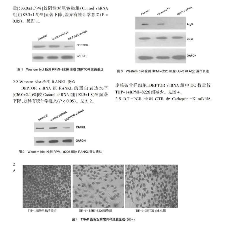
Western blot检测显示,慢病毒感染RPMI-8226细胞72 h后,DEPTOR shRNA组DEPTOR蛋白表达量[(33.0±1.7)%]较阴性对照转染组(Control shRNA组)[(89.3±1.5)%]显著下降,差异有统计学意义(P < 0.05)。见图1。
2.2 Western blot检测RANKL蛋白
DEPTOR shRNA组RANKL的蛋白表达水平[(36.0±2.1)%]较Control shRNA组[(92.5±1.8)%]显著下降,差异有统计学意义(P < 0.05)。见图2。
2.3 Western blot技术检测自噬相关蛋白LC-3和Atg5
DEPTOR shRNA组Atg5蛋白[(18.6±2.1)%]表达量与Control shRNA组[(48.5±1.8)%]相比显著下降,DEPTOR shRNA组LC-3Ⅱ的蛋白表达水平[(11.3±1.6)%]与Control shRNA组[(22.6±1.9)%]相比下调,差异均有统计学意义(P < 0.05)。见图3。
2.4 DEPTOR shRNA对破骨样细胞生成的影响
本研究显示,THP-1细胞单培养组TRAP染色呈阴性;THP-1+RPMI-8226组中可见TRAP染色阳性多核破骨样细胞,DEPTOR shRNA组中OC数量较THP-1+RPMI-8226组减少。见图4。
2.5 RT-PCR检测CTR和Cathepsin-K mRNA表达
RT-PCR检测显示,THP-1+RPMI-8226细胞组中CTR和Cathepsin-K基因表达水平均较THP-1细胞单培养组升高(P < 0.05);THP-1+DEPTOR shRNA组中CTR和Cathepsin-K基因表达水平较THP-1+RPMI-8226细胞组均降低(P < 0.05);THP-1+DEPTOR shRNA组中CTR和Cathepsin-K基因表达水平与THP-1细胞单培养组比较差异无统计学意义(P > 0.05)。见图5。
3 讨论
人外周血中的OC前体细胞,在M-CSF与RANKL的共同刺激下,可分化为OC[11]。M-CSF能够与OC前体细胞表面的c-Fms受体结合,并维持OC前体细胞的生存。RANKL通过激活OC前体细胞上的受体使其分化成OC,并增强OC活力。OC与MM细胞相互作用所导致的恶性循环是MBD发生进行性骨质破坏最主要的原因[12]。
自噬具有维持细胞自稳的功能。近年来研究表明,细胞自噬与发育、氧化性损伤保护、神经退行性疾病及肿瘤细胞的增殖有关,并且多种人类肿瘤细胞中存在自噬活性的改变[13],发现在MM细胞中存在较高的自噬活性[14],调节自噬可能会为MM治疗提供新角度。目前研究显示DEPTOR-mTOR信号通路在调控细胞生长、分化、凋亡、自噬等活动中起重要作用[15-16]。位于mTOR信号通路下游的核糖体蛋白p70S6K抑制细胞自噬发生,其活性受mTOR调节。有研究发现自噬活性改变参与了骨髓间充质干细胞向成骨细胞的分化过程,研究也提示自噬参与了K562细胞向巨核细胞的分化演变进程[17]。
自噬和OC功能关系密切,有研究发现OC产生自噬可能是OC的一种生存保护机制,自噬对机体骨形成及骨骼发育有重要调节作用[18],但相关分子机制并不清楚。目前发现,自噬主要受mTOR信号通路的调节,DEPTOR是mTOR的负性因子,在MM细胞株中DEPTOR基因沉默后下调自噬[9]。调节自噬活性可能对MM微环境下OC分化有重要影响,从而可通过调节自噬阻断OC-MM恶性循环。
本研究采用RNA干扰技术,沉默DEPTOR基因,研究了在非接触式共培养模式下观察OC分化的过程。目前已知PMA刺激THP-1可使其分化为贴壁的巨噬细胞,在RANKL和M-CSF诱导下,可分化形成TRAP染色陽性的OC[19]。本研究显示,MM细胞RPMI-8226可诱导THP-1细胞破骨样分化,形成TRAP阳性多核破骨样细胞,OC特征性基因CTR和Cathepsin-K的mRNA表达也增高。
本研究表明沉默DEPTOR蛋白表达后可明显下调RPMI-8226细胞RANKL蛋白表达。沉默DEPTOR可抑制细胞的自噬功能,MM细胞株可表达RANKL。而RANKL有诱导单核巨噬细胞系分化为OC的功能。本研究显示DEPTOR shRNA能明显抑制共培养体系中THP-1细胞的破骨样分化,该作用可能与DEPTOR下调RPMI-8226细胞RANKL有关,DEPTOR-mTOR信号通路参与的自噬可能在RANKL介导的OC分化过程中发挥重要影响,抑制自噬可阻碍OC的分化成熟。
[参考文献]
[1] Gay F,Palumbo A. Management of older patients with mul?鄄tiple myeloma [J]. Blood Rev,2011,25(2):65-73.
[2] Edwards JR,Weivoda MM. Osteoclasts:malefactors of disease and targets for treatment [J]. Discov Med,2012,13(70):201-210.
[3] McManus S,Bisson M,Chamberland R,et al. Autophagy and 3-Phosphoinositide-Dependent Kinase 1(PDK1)-Rel?鄄ated Kinome in Pagetic Osteoclasts [J]. J Bone Miner Res,2016,31(7):1334-1343.
[4] Owen HC,Vanhees I,Gunst J,et al. Critical illness-induced bone loss is related to deficient autophagy and histone hypomethylation [J]. Intensive Care Med Exp,2015,3(1):52.
[5] Gómez-Puerto MC,Verhagen LP,Braat AK,et al. Activation of autophagy by FOXO3 regulates redox homeostasis during osteogenic differentiation [J]. Autophagy,2016,12(10):1804-1816.
[6] Puissant A,Robert G,Auberger P. Targeting autophagy to fight hematopoietic malignancies [J]. Cell Cycle,2010,9(17):3470-3478.
[7] Hadji P,Coleman R,Gnant M. Bone effects of mammalian target of rapamycin(mTOR)inhibition with everolimus [J]. Critical Reviews in Oncology/Hematology,2013,87(2):101-111.
[8] Peterson TR,Laplante M,Thoreen CC,et al. DEPTOR is an mTOR inhibitor frequently overexpressed in multiple myeloma cells and required for their survival [J]. Cell,2009, 137(5):873-886.
[9] Zhang H,Chen J,Zeng Z,et al. Knockdown of DEPTOR induces apoptosis,increases chemosensitivity to doxorubicin and suppresses autophagy in RPMI-8226 human multiple myeloma cells in vitro [J]. Int J Mol Med,2013, 31(5):1127-1134.
[10] 张浩然,曾志勇,陈君敏.体外RNA干扰下调DEPTOR表达对人多发性骨髓瘤细胞增殖和凋亡能力的影响[J].中国生物工程杂志,2013,33(5):13~21.
[11] S?rensen MG,Heiksen K,Schaller S,et al. Characterization of osteoclasts derived from CD14+ monocytes isolated from peripheral blood [J]. J Bone Miner Metab,2007, 25(1):36-45.
[12] Fowler JA,Edwards CM,Croucher PI. Tumor-host cell interactions in the bone disease of myeloma [J]. Bone,2011,48(1):121-128.
[13] Mizushima N. Autophagy:process and function [J]. Genes Dev,2007,21(22):2861- 2873.
[14] Hoang B,Benavides A,Shi Y,et al. Effect of autophagy on multiple myeloma cell viability [J]. Mol Cancer Ther,2009,8(7):1974-1984.
[15] Yang Z,Klionsky DJ. Mammalian autophagy:core molec?鄄ular machinery and signaling regulation [J]. Curr Opin Cell Biol,2010,22(2):124-131.
[16] 黃漫华,葛鸿庆,陈文治,等.葛根素诱导破骨细胞自噬的机制研究[J].中国医药导报,2015,12(15):20-23.
[17] Colosetti P,Puissant A,Robert G,et al. Autophagy is an important event for megakaryocytic differentiation of the chronic myelogenous leukemia K562 cell line [J]. Autop?鄄hagy,2009,5(8):1092-1098.
[18] Usategui-Martín R,García-Aparicio J,Corral-Gudino L,et al. Polymorphisms in autophagy genes are associated with paget disease of bone [J]. PLoS One,2015,10(6):e0128984.
[19] Munoz-Pacheco P,Ortega-Hernandez A,Miana M,et al. Ezetimibe inhibits PMA-induced monocyte/macrophage differentiation by altering microRNA expression:a novel anti-atherosclerotic mechanism [J]. Pharmacol Res,2012, 66(6):536-543.
(收稿日期:2017-11-07 本文编辑:任 念)